Next: Conclusions
Up: Sava and Biondi: WEMVA
Previous: Wave-equation migration velocity analysis
In our first example, we consider a background wavefield
emerging from
a fixed point source on the surface, but investigate
the sensitivity kernels for various points in the image.
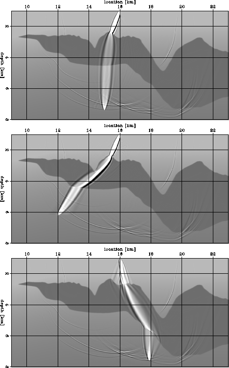
Each panel in Figure 1 depicts a superposition of three elements:
the velocity model, the band-limited wavefield corresponding to a point
source on the surface, and the sensitivity kernel corresponding to a point in the subsurface.
A fundamental problem with ray-based
MVA is that rays are poor approximations
of the actual wavepaths when a band-limited
seismic wave propagates through a rugose
top of the salt.
Figure 1 illustrates this issue quite clearly.
It shows three sensitivity kernels for frequencies of 1-26 Hz.
The top panel in Figure 1 shows a wavepath
that could be reasonably approximated using the method introduced by
Lomax (1994) to trace fat rays using asymptotic methods.
In contrast,
the wavepaths shown in both the middle and bottom panels
cannot be well approximated using Lomax' method.
The amplitude and shapes of these wavepaths are much more complex
than a simple fattening of a geometrical ray could ever describe.
The bottom panel illustrates the worst
situation for ray-based tomography
because the rugosity of the top of the
salt has the same scale as the spatial wavelength of the seismic wave.
zifat
Figure 1 Kinematic sensitivity kernels for frequencies between 1 and 26 Hz
for various locations in the image and a
point on the surface.
Each panel is an overlay of three elements:
the slowness model, the wavefield corresponding
to a point source on the surface at x=16 km, and
wave paths (sensitivity kernels) from a point in the subsurface to the
source.
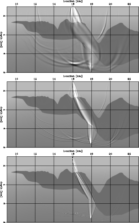
 zifrq2
Figure 2 Frequency dependence of kinematic sensitivity kernels between a
location in the image and a point on the surface.
Each panel is an overlay of three elements:
the slowness model, the wavefield corresponding
to a point source on the surface at x=16 km, and
wave paths (sensitivity kernels) from a point in the subsurface to the
source.
The different wave paths correspond to frequency bands of
1-5 Hz (top),
1-16 Hz (middle) and
1-64 Hz (bottom).
The larger the frequency band, the narrower the wave path.
The end member for an infinitely wide frequency band
corresponds to an infinitely thin geometrical ray.
The fundamental reason why the true wavepaths cannot be approximated
using fattened geometrical rays is that
they are frequency dependent.
Figure 2 illustrates this dependency
by depicting the wavepath shown in the bottom panel of
Figure 1 as a function
of the temporal bandwidth:
1-5 Hz (top), 1-16 Hz (middle), and
1-64 Hz (bottom).
The width of the wavepath decreases
as the frequency bandwidth increases,
and the focusing and defocussing of the
energy varies with the frequency bandwidth.
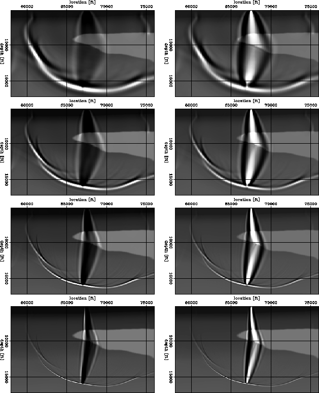
In the next example
(Figures 3 and 4)
we compare the shapes of sensitivity kernels
when we change the type of source for the background wavefield,
its frequency content and the method used to generate an image
perturbation in the subsurface. As for the preceding example,
we show the results as a superposition of the velocity model,
the background wavefield and the sensitivity kernel from a
fixed point in the subsurface.
Figure 3 shows the sensitivity kernels for a point source on the
surface, and
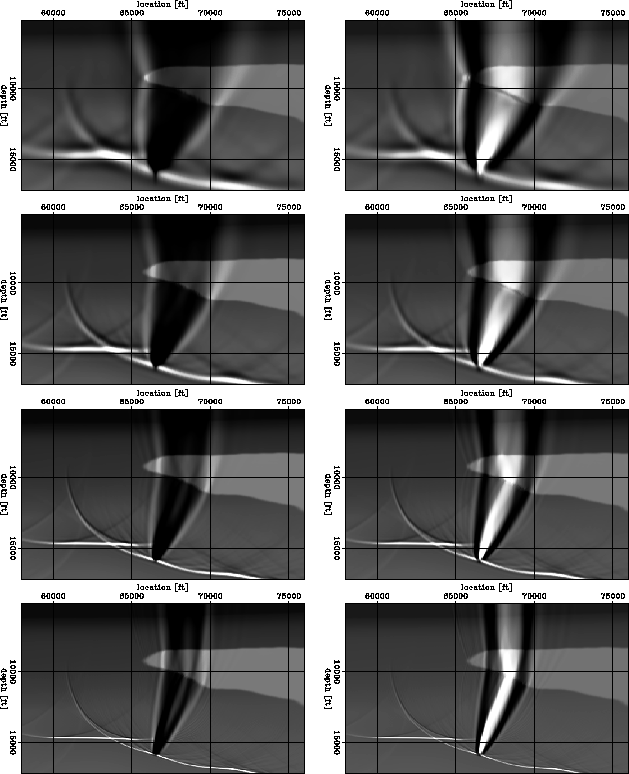
Figure 4 shows the sensitivity kernels for a plane-wave propagating
vertically at the surface.
In both pictures, the left column corresponds to kinematic
image perturbations of equation (6), and the
right column corresponds to amplitude image perturbations
of equation (7) obtained by scaling of the background image
by an arbitrary number.
From top to bottom, we show sensitivity kernels of increasing frequency range:
1-4 Hz, 1-8 Hz, 1-16 Hz and 1-32 Hz.
Once again, we can see the large frequency dependence of the
sensitivity kernels. The area of sensitivity reduces with
increased frequency which is a clear indication that
a frequency dependent migration velocity analysis method
like WEMVA can better handle subsalt environments with
patchy illumination and that illumination itself is a frequency
dependent phenomenon which needs to be addressed in this way.
fat2d.Tray2a
Figure 3 The dependence of sensitivity kernels to frequency and image perturbation.
From top to bottom, the frequency range is
1-4 Hz, 1-8 Hz, 1-16 Hz and 1-32 Hz.
The left column corresponds to kinematic image perturbations,
and the right column corresponds to dynamic image perturbations.
The wavefield is produced from a point source.
![[*]](http://sepwww.stanford.edu/latex2html/movie.gif) fat2d.Tray2b
fat2d.Tray2b
Figure 4 The dependence of sensitivity kernels to frequency and image perturbation.
From top to bottom, the frequency range is
1-4 Hz, 1-8 Hz, 1-16 Hz and 1-32 Hz.
The left column corresponds to kinematic image perturbations,
and the right column corresponds to dynamic image perturbations.
The wavefield is produced by a horizontal incident plane-wave.
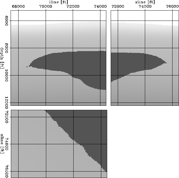
Finally, we show wave-equation MVA sensitivity kernels
for a 3D velocity model Figure 5
corresponding to a salt environment.
We consider the case of a point source on the
surface and data with a frequency range of
1-16 Hz.
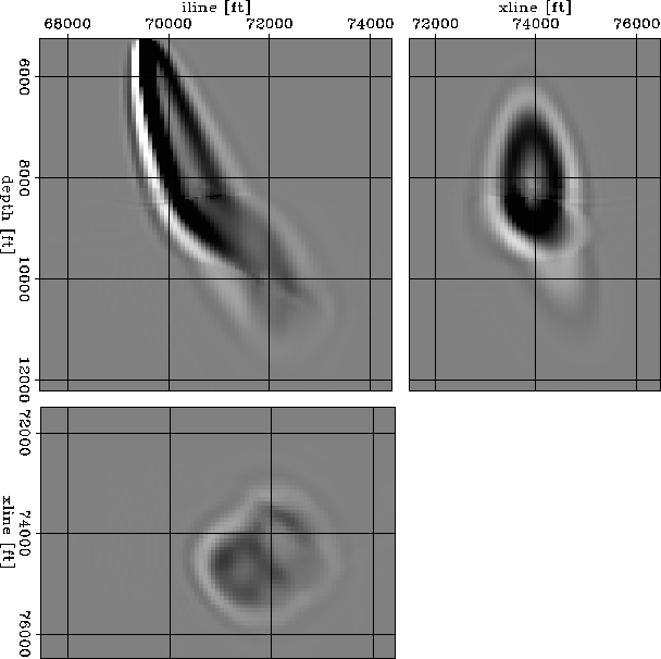
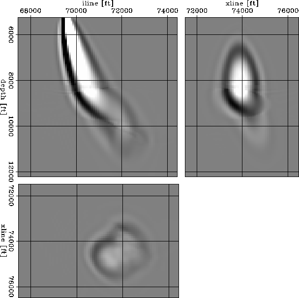
Figure 6 shows the sensitivity kernel for a kinematic
image perturbation, while Figure 7 for
a amplitude image perturbation.
In both cases, the shapes of the kernels are
complicated, which is an expression of the
multipathing occurring as waves propagate
through rough salt bodies.
The horizontal slice
indicates multiple paths
linking the source point on the surface with
the image perturbation in the subsurface.
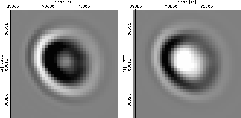
One noticeable characteristic is that the sensitivity kernels
constructed from amplitude image perturbations
show the largest sensitivity in the center of the
kernel, as opposed the kinematic kernels which
show the largest sensitivity away from the central path.
This phenomenon was discussed by
Dahlen et al. (2000) in the
context of finite-frequency traveltime tomography.
We illustrate it for WEMVA in Figure 8 with two
horizontal slices
in the sensitivity kernels shown in Figures 6 and
7.
fat3.sC
Figure 5 3D slowness model.
fat3.fp3
Figure 6 3D sensitivity kernels for wave-equation MVA.
The frequency range is 1-16 Hz.
The kernels are complicated by the multipathing
occurring as waves propagate through the rough
salt body.
The image perturbation corresponds to a kinematic shift.
fat3.fq3
Figure 7 3D sensitivity kernels for wave-equation MVA.
The frequency range is 1-16 Hz.
The kernels are complicated by the multipathing
occurring as waves propagate through the rough
salt body.
The image perturbation corresponds to an amplitude scaling.
fat3.svty
Figure 8 Cross-section of 3D sensitivity kernels for wave-equation MVA.
The left panel corresponds to an image perturbation
produced a kinematic shift, while the right panel
corresponds to an image perturbation produced by
amplitude scaling.
The lowest sensitivity occurs in the center of the
kinematic kernel (left). In contrast, the maximum
sensitivity occurs in the center of the kernel (right).
Next: Conclusions
Up: Sava and Biondi: WEMVA
Previous: Wave-equation migration velocity analysis
Stanford Exploration Project
5/23/2004