


![[*]](http://sepwww.stanford.edu/latex2html/cross_ref_motif.gif) we found the Sea of Galilee
in Figure to be too smooth for viewing pleasure
so we made the roughened versions
in Figure
using a filter based
on equation (),
a one-dimensional filter that we could apply
over the x-axis or the y-axis.
In Fourier space such a filter has a response function of kx
or a function of ky.
The isotropy of physical space tells us
it would be more logical to design a filter that
is a function of
kx2+ky2.
In Figure we saw that the helix derivative
we found the Sea of Galilee
in Figure to be too smooth for viewing pleasure
so we made the roughened versions
in Figure
using a filter based
on equation (),
a one-dimensional filter that we could apply
over the x-axis or the y-axis.
In Fourier space such a filter has a response function of kx
or a function of ky.
The isotropy of physical space tells us
it would be more logical to design a filter that
is a function of
kx2+ky2.
In Figure we saw that the helix derivative
It is nice having the 2-D helix derivative,
but we can imagine even nicer 2-D low-cut filters.
In one dimension,
equation
()
and
()
we designed a filters with an adjustable parameter,
a cutoff frequency.
We don't have such an object in 2-D so
I set out to define one.
It came out somewhat
abstract and complicated,
and didn't work very well,
but along the way
I found a simpler parameter that is very effective in practice.
We'll look at it first.
|
mam
Figure 10 Mammogram (medical X-ray). The cancer is the ``spoked wheel.'' (I apologize for the inability of paper publishing technology to exhibit a clear grey image.) The white circles are metal foil used for navigation. The little halo around a circle exhibits the impulse response of the helix derivative. | 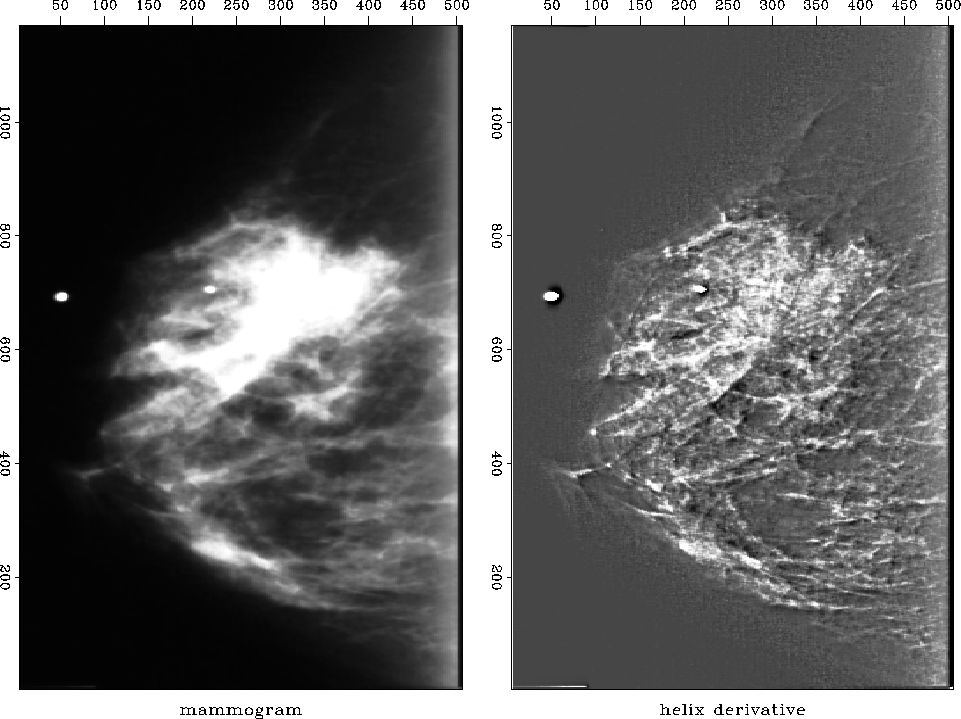 |
![[*]](http://sepwww.stanford.edu/latex2html/movie.gif)


First I had a problem preparing Figure .
It shows shows the application of the helix derivative
to a medical X-ray.
The problem was that the original X-ray was all positive
values of brightness so there was a massive amount of
spatial low frequency present.
Obviously an x-derivative or a y-derivative would
eliminate the low frequency, but the helix derivative did not.
This unpleasant surprise arises
because the filter in equation
(11)
was truncated after a finite number of terms.
Adding up the terms actually displayed in equation
(11),
they sum to .183 whereas theoretically the sum of all the terms should be zero.
From the ratio of .183/1.791 we can say that the filter
pushes zero frequency amplitude 90% of the way to zero value.
When the image contains very much zero frequency amplitude,
this is not good enough.
Better results could be obtained with more coefficients,
and I did use more coefficients,
but simply removing the mean saved me
from needing a costly number of filter coefficients.
We can visualize a plot of the magnitude of the 2-D
Fourier transform of the filter
(11).
It is a 2-D function of kx and ky and it should
resemble ![]() .It does look like this even when the filter
(11)
has been truncated.
The point of the cone
.It does look like this even when the filter
(11)
has been truncated.
The point of the cone ![]() becomes
rounded and the truncated approximation of
kr does not reach zero at the origin of the (kx,ky)-plane.
We can force it to vanish at zero frequency
by subtracting .183 from the lead coefficient 1.791.
I did not do that subtraction in Figure
which explains the whiteness in the middle of the lake.
becomes
rounded and the truncated approximation of
kr does not reach zero at the origin of the (kx,ky)-plane.
We can force it to vanish at zero frequency
by subtracting .183 from the lead coefficient 1.791.
I did not do that subtraction in Figure
which explains the whiteness in the middle of the lake.
 |
Now let us return to my more logical but less effective approach.
I prepared a half dozen medical X-rays like Figure
.
The doctor brought her young son to my office one evening
to evaluate the results.
In a dark room I would show the original X-ray on a big screen
and then suddenly switch to the helix derivative.
Every time I did this, her son would exclaim ``Wow!''
The doctor was not so easily impressed, however.
She was not accustomed to the unfamiliar image.
Fundamentally, the helix derivative applied to her data
does compress the dynamic range making weaker features more readily discernable.
We were sure of this from theory and from
various geophysical examples.
The subjective problem was her unfamiliarity with our display.
I found that I could always spot anomalies more quickly
on the filtered display, but then I would feel more comfortable
when I would discover those same anomalies also present
(though less evident) in the original data.
Thinking this through, I decided the doctor would likely have
been more impressed
had I used a spatial lowpass filter instead of the helix derivative.
That would have left the details of her image
(above the cutoff frequency)
unchanged
altering
only the low frequencies,
thereby allowing me to increase the gain.
In 1-D we easily make a low-cut filter
by compounding a first derivative (which destroys low frequencies)
with a leaky integration (which undoes the derivative at all other frequencies).
We can do likewise with a second derivative.
In Z-transform notation, we would use something like
(-Z-1 + 2.00 - Z) / (-Z-1 + 2.01 - Z).
(The numerical choice of the .01 controls the cutoff frequency.)
We could use spectral factorization to break this spectrum into
causal and anticausal factors.
The analogous filter in 2-D is
![]() which could also be factored as we did the helix derivative.
I tried it.
I ran into the problem that my helix derivative operator
had a practical built-in parameter, the number of coefficients,
which also behaves like a cutoff frequency.
If I were to continue this project,
I would use expressions for
which could also be factored as we did the helix derivative.
I tried it.
I ran into the problem that my helix derivative operator
had a practical built-in parameter, the number of coefficients,
which also behaves like a cutoff frequency.
If I were to continue this project,
I would use expressions for
![]() directly in the Fourier domain where there is only one adjustable parameter,
the cutoff frequency k0,
and there is no filter length to confuse the issue and puff-up the costs.
directly in the Fourier domain where there is only one adjustable parameter,
the cutoff frequency k0,
and there is no filter length to confuse the issue and puff-up the costs.
A final word about the doctor. As she was about to leave my office she suddenly asked whether I had scratched one of her X-rays. We were looking at the helix derivative and it did seem to show a big scratch. What should have been a line was broken into a string of dots. I apologized in advance and handed her the original film negatives which she proceeded to inspect. ``Oh,'' she said, ``Bad news. There are calcification nodules along the ducts.'' So the scratch was not a scratch, but an important detail that had not been noticed on the original X-ray.
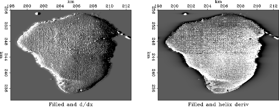
In preparing an illustration for here,
I learned one more lesson.
The scratch was small,
so I enlarged a small portion of the mammogram for display.
The very process of selecting a small portion
followed by scaling the amplitude
between maximum and minimum darkness of printer ink
had the effect enhancing the visibility of the scratch on
the mammogram itself.
Now Figure shows it to be
perhaps even clearer than on the helix derivative.
 |
An operator for applying the helix filter is
helderiv .
helderivhelix-derivative filter