

![[*]](http://sepwww.stanford.edu/latex2html/prev_gr.gif)


Geophysical experience with helix derivative. One day a group of us were scanning 300 images of seismic reflectivity looking for about 50 river meanders buried beneath the Gulf of Mexico. We had side-by-side views of an original and a helix derivative. Independently, each of us found ourself looking first at the helix derivative, because it was easier to look at, and then looking back to the original to see if we were deceived. Only on rare occasions was there much difference. Conclusion: The derivative is best for the first look. Things can be overlooked on the original that are not overlooked on the helix. On careful study, the difference is less.
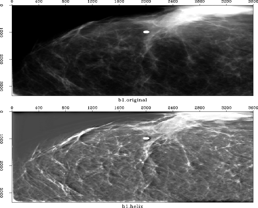
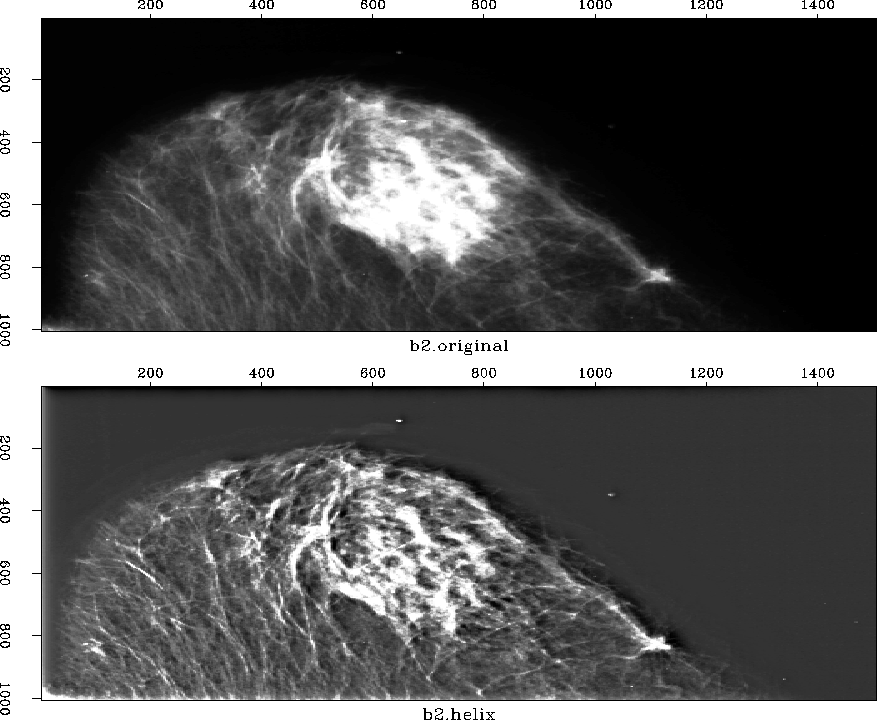
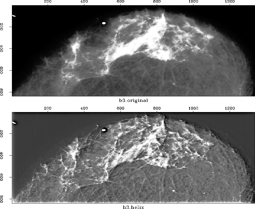
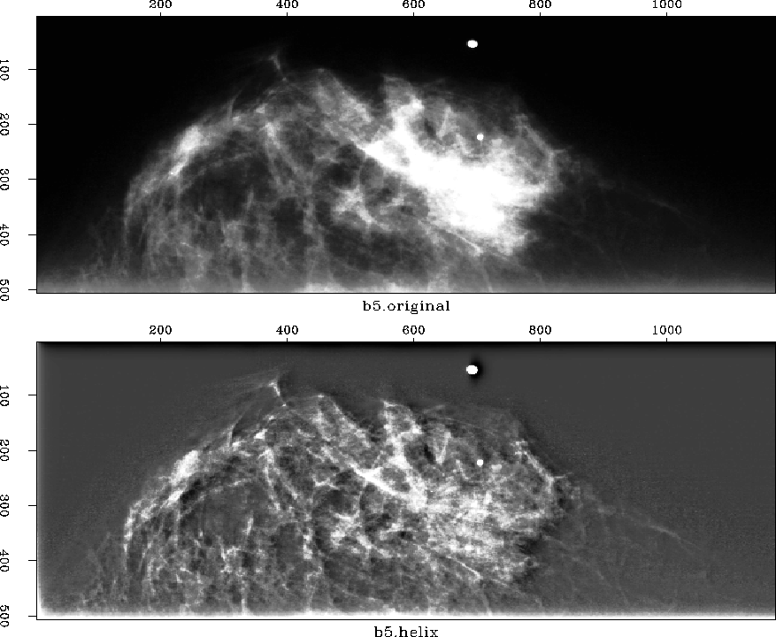
Mammogram processing. Below is the result of applying my helical derivative process to some mammograms. There are several minor aspects of the processing that I could play with. One is the mean balance. The original maps the all-positive signal to the full color pallet from black to white. My helicon process produces some negatives, and therefore I have shifted the color pallet to see them. I could shift by various amounts and don't know which is best since I don't know what the doctor is looking for.
Simple explanation of my process. The process that I invented and call helix derivative has a simple explanation. Tiny white areas remain white. Tiny black areas remain black. Larger areas (both black and white) are pushed towards gray. A mathematical consequence of this process is that a white dot or line acquires a faint black shadow. This shadow speeds human recognition of the dot or line, but it also creates an image that is in some sense false. My recommendation is to put both images in a film loop and run the loop slowly, about one frame per second.
Results and interpretation. In figure 1, I notice that the nature of the white mass is quite homogeneous on the original. On the helix derivative, we see that the white mass is "somewhat spotty" on the left and "more homogeneous" on the right. Generally, viewing in the lab is much better than viewing over the web. In the lab we have higher resolution and we can quickly blink back and forth between the two images which enables better correlations. With geophysical images, and this one too, we often find that we notice features more quickly on the helix derivative, but after noticing them, we can usually see them also in the original.
 |


 |
 |
 |
 |
REFERENCE
Medical links. Here is a mammogram of a fibroadenoma . I don't know if this is what the doctor is looking at. Here is another one , some calcifications , a medical tutorial in North Carolina.
Digital mammography links. There is an image database in Nijmegen, Holland. Some research at Livermore . There are many links at Brandise U . Stanford University ISL is next door. The best digital-mammography data-processing research site I found is at Columbia University . They use wavelet theory and Java but I find their server speed quite slow.